What is Knee Cap Dislocation (Patella Dislocation)?
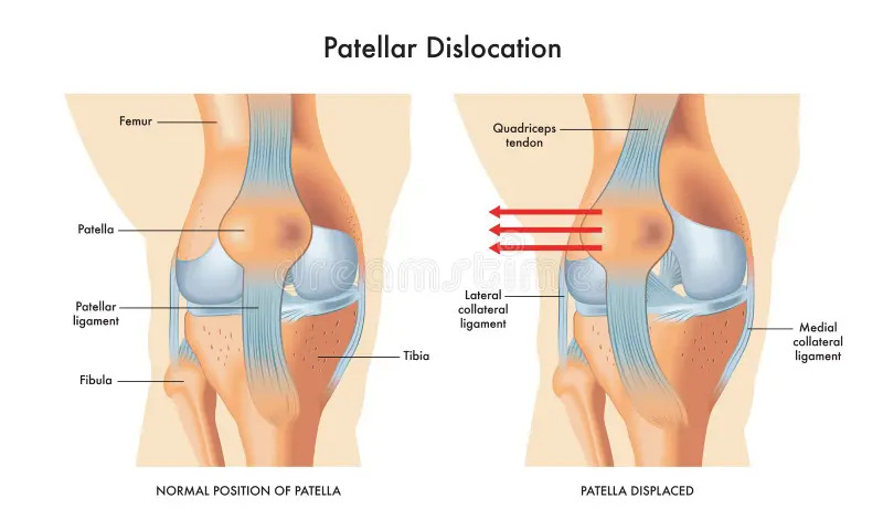
A Knee Cap Dislocation (Patella Dislocation) means that the kneecap is completely out of the groove on the thigh (femur) bone and is outside of the knee joint. In general, a severe trauma or impact is required for the kneecap to dislodge the first time, while recurrent dislocations occur much more easily.
The joint between the kneecap and the thigh bone is called the patellofemoral joint. The cartilage layer under the kneecap and the hollow part of the thigh bone form the faces of the joint. The presence of the kneecap allows the quadriceps muscle, which allows us to open and lock our knee on the front of the thigh, to work more efficiently. Various ligaments and muscles hold the kneecap where it should be.
The connective tissue tensor fascia lata, patellar ligament, knee joint capsule, patellofemoral ligament, meniscopatellar ligament, quadriceps muscle, which descends from the hip to the knee on the outer side of the thigh, are the structures that hold the kneecap.
Kneecap dislocation is most commonly outward. Meanwhile, the inner muscles and ligaments are overstretched and damaged. Partial displacement of the kneecap without complete removal is called subluxation.
Knee Cap Dislocation (Patella Dislocation) occurs in two ways.
These ;
- Acute (Initial) Patella Dislocation
- Chronic (Recurrent) Patella Dislocation
What is Acute (Initial) Knee Cap Dislocation (Patella Dislocation) ?
There is usually a trauma in the first (acute) patellar dislocation. During this trauma, the MPFL (Medial Patellofemoral Ligament) ligament, which is the most important supporter of the patella, breaks.
The patella slides outward and comes out. Most of the time, the patella that protrudes outwards fits back to the trochlea with knee movement. But now the MPFL ligament, which keeps the patella in balance on the trochlea, is broken. Therefore, there is a risk of recurrence.
What is Chronic (Recurrent) Knee Cap Dislocation (Patella Dislocation)?
It is the outward sliding of the patella with simple knee movements or sudden movements without trauma. This situation recurs frequently.
What are the Causes of Recurrence of Knee Cap Dislocation (Patella Dislocation) ?
The MPFL ligament that was broken during the first dislocation has not healed.
Congenital or acquired anatomical differences.
For example;
- The pit of the trochlea, which we call the nest of the patella, is shallow or flat, that is, the pit is not formed (Trochlear Dysplasia). In this case, the patella cannot sit in the socket and slides out easily.
- The patella is located higher than the normal location (Patella Alta). In this case, since the patella is not over the trochlear socket, it easily slides outward.
- Attachment of the patellar tendon, which connects the patella to the tibia, to the tibia more externally than normal. In this case, the kneecap is pulled outward by the patellar tendon and forced to protrude.
- The MPFL ligament is longer and more flexible than normal. In this case, this ligament, which is the most important supporter that keeps the patella in its socket, cannot work.
- Some structural or later anatomical defects in the thighbone (femur) and tibia (tibia) also cause patellar dislocation.
What are the Causes of Knee Cap Dislocation (Patella Dislocation)?
If the knee joint is turned inward in a slightly bent position while the foot is flat on the floor, the kneecap may come out. This usually happens during exercise and is called an acute or traumatic dislocation. Typical sports that can cause this injury are football, handball, dance and gymnastics.
Acute patellar dislocation is the most common form with 80 percent. Patella dislocation is less common. In the case of subluxation, the kneecap moves sideways in its socket without protruding completely. This can happen due to a previous knee injury or ligaments that are too loose.
What are the Symptoms of Knee Cap Dislocation (Patella Dislocation)?
People with this disorder say that the kneecap moves too much, especially when folding the knees while walking, climbing stairs or standing up from a sitting position, or in the same way, if they climbed too many stairs or walked too much, the knee hurts or even swells, and even heats up. It causes serious knee pain by affecting the quality of life of people too much.
If no precautions are taken, it will force the ligaments around the kneecap and cause ligament injuries in the future.
Another question that comes to mind is why does this kneecap tend to protrude? The reason for this is that the muscles in the outer group of our leg are stronger than the muscles in the inner group, and in case of weakening, the muscles in the outer group of the leg pull the kneecap out more, causing it to come out more than it should be. This is an indication of knee slippage, that is, patellar dislocation.
How is Knee Cap Dislocation (Patella Dislocation) Diagnosed?
The diagnosis of patellar dislocation is made by the patient’s history, detailed physical examination, direct radiographs, MRI and, if necessary, CT (computed tomography). Apart from making a diagnosis, it is necessary to examine the cause of the patient’s patella dislocation and all the underlying anatomical differences. Correct treatment is possible only by detecting all anatomical problems and correcting the necessary ones.
What are the Risk Factors for Knee Cap Dislocation (Patella Dislocation)?
There are many factors that increase the risk of kneecap dislocation.
These include:
- Deformities in legs such as crooked legs,
- Deformed locomotion or other anatomical features,
- The kneecap is higher than normal,
- A weak inner thigh muscle,
- Hypermobile (overly mobile) joints or weak ligaments,
How is Knee Cap Dislocation (Patella Dislocation) Treated?
The first treatment is to replace the kneecap. If it doesn’t fit on its own, don’t try to force it, go to the emergency room or orthopedist.
Treatment methods according to the degree of the disease are as follows:
- Exercise,
- Knee corset,
- Physical therapy or surgery is recommended.
If the patient applied in the early period, pain-relieving and cartilage-strengthening medication and exercise program are recommended.
In advanced cases, the kneecap is placed in place with arthroscopic surgery and the worn tissues are cleaned.
Depending on the condition of the surgery, the patient can return to his daily life after 3 to 4 weeks. Knee prosthesis can also be applied when necessary.
