What is Retinopathy of Prematurity (ROP)?
Retinopathy of Prematurity (ROP) examination is a method used by the ophthalmologist to examine the retina (layer of the eye) of premature babies in detail. This examination is performed in cases where babies have risk factors such as premature birth and low birth weight.
Retinopathy of Prematurity (ROP) examination is important to early diagnose any abnormalities that may occur in the baby’s eye and start treatment if necessary. Rope examination is usually performed between 4 and 9 weeks after birth. The baby is anesthetized with eye drops and the ophthalmologist examines the baby’s eye using a special instrument.
During this examination, symptoms such as abnormalities that may occur in the retina, vascular enlargement or bleeding are looked for. Early diagnosis is vital to protect the baby’s eye health and ensure treatment of progressive conditions.
Rope examination is of great importance for the development of vision abilities of premature babies. Babies born prematurely do not have enough time for normal vascular growth in the retina to complete. Therefore, early diagnosis and treatment of ROP plays a critical role in preventing or reducing vision loss.
In order to protect the eye health of babies, it is important to perform FRO examination regularly and to intervene quickly in cases that require treatment. Contacting a specialist ophthalmologist is the best step to monitor the baby’s eye health and apply appropriate treatments when necessary.
How Does Retinopathy of Prematurity (ROP) Occur?
The birth of the baby before the vascular development of the retina is completed causes the wrong vascularization to appear in these avascular areas due to wrong stimulation (VEGF release). These wrong vessels cause bleeding within the eye and separation of the nerve layer (Retinal Detachment), leading to vision loss.
The risk of ROP increases as birth weight and gestational age decrease. While the rate of premature retinopathy is almost 80 percent in a 25-week-old baby, this rate is around 10 percent in a baby under 32 weeks. In 10 percent of cases, it may regress spontaneously and recover without the need for treatment. However, controls are continued until vascularization is completed.
ROP can also be seen in babies born prematurely (36th week) in addition to babies weighing 1500 grams and under 32 weeks. If systemic risk factors are very prominent, the neonatologist may still recommend ROP examination. However, all premature babies weighing 1500 grams and before the 32nd week should be examined for ROP.
What are The Causes of Retinopathy of Prematurity (ROP)?
The most important causes of retinopathy of prematurity include premature birth and low birth weight. In a normal pregnancy, vascular development of the retina in the eyes of babies begins from the 16th week of pregnancy and is usually completed by the 40th week.
However, in cases of prematurity, that is, when babies are born early, this vascularization may not be completed yet. Babies born prematurely often experience systemic problems such as respiratory problems.
In response to breathing problems, these babies are given high doses of oxygen therapy. Administration of high doses of oxygen therapy is one of the main risk factors in the development of retinopathy of prematurity.
Additionally, the presence of factors such as anemia, intracranial hemorrhages, and congenital anomalies may also increase the risk of retinopathy of prematurity.
What Are The Symptoms of Retinopathy of Prematurity (ROP)?
ROP has no symptoms visible to the naked eye. In advanced cases of ROP, the retina may partially or completely move away from its normal position at the back of the eye. This is called retinal detachment and can cause vision loss and blindness.
If your baby has ROP that causes damage, you may later notice:
- Their eyes may wander, tremble, or make other unusual movements.
- Their eyes may not follow objects.
- Pupils may appear white.
- May have difficulty recognizing faces.
Babies with ROP are more likely to have other eye problems as they get older,
For example:
- Retinal Detachment
- Myopia
- Amblyopia (Lazy Eye)
- Strabismus
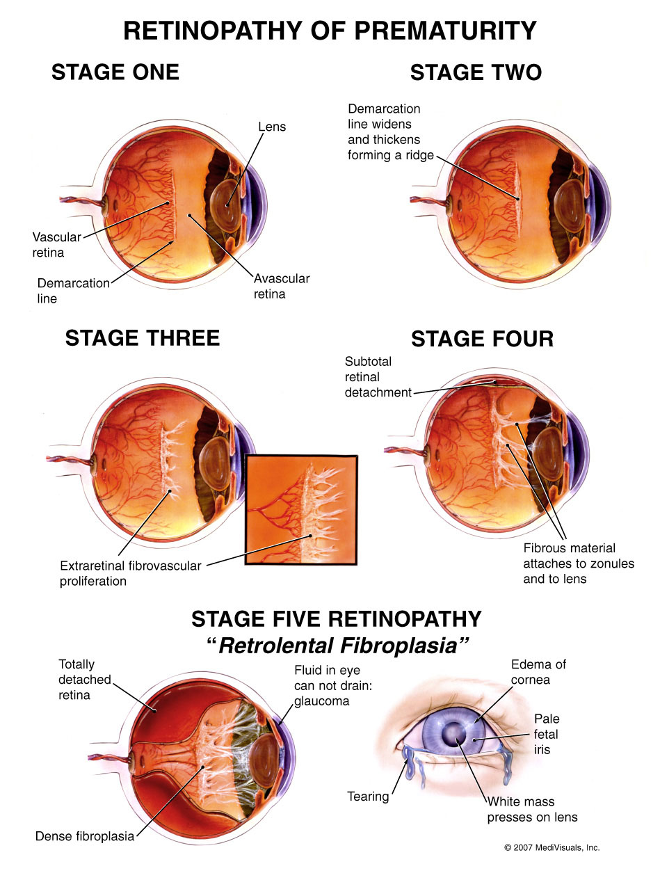
Retinopathy of Prematurity (ROP) Stages and Classification of the Disease
The areas used to classify the disease are as follows:
Zone 1:
Circular area with a diameter twice the distance between the macular region and the optic disc, with the optic disc in the middle region.
Zone 2:
Circular area with the optic disc at its center and containing the entire retina downwards
Zone 3:
Crescent-shaped area outside zone 2 and in the opposite direction of this zone.
The disease is discussed in 5 different stages and these stages are as follows:
Stage 1:
It is the phase where abnormal and slight growth occurs in blood vessels.
Stage 2:
Moderate abnormal vascular enlargement.
The disease may improve and regress on its own in this 2nd stage, or it may develop into the 3rd stage after this stage.
Stage 3:
High-speed, abnormal vascular growth in the retina.
Stage 4:
Beginning of detachment in the retina.
Stage 5:
The final stage of the disease and retinal detachment occurs.
Who is at Risk and Should Be Followed?
- Those born at 32 weeks and earlier
- Babies weighing 1500 grams and less
- Those who have received intensive oxygen therapy
- Babies with other accompanying diseases
- Babies considered risky by the pediatrician
How is Retinopathy of Prematurity (ROP) Diagnosed?
The diagnosis of Retinopathy of Prematurity is made by examination. Babies with low gestational age and low birth weight are referred for an eye examination by the neonatal doctor 28 days after birth.
After the initial examination, the ophthalmologist examines the retina at the back of the eye indirectly with an ophthalmoscope and determines the frequency of follow-up according to the ROP finding and its severity. If there is no risk of ROP, the patient is monitored every two weeks until vascularization is completed.
However, if signs of ROP develop, depending on the severity and stage of the symptom, it is closely monitored once a week or even every 2-3 days, and emergency treatment is applied when necessary.
How is Retinopathy of Prematurity (ROP) Examined?
This examination, which takes approximately 10-15 minutes, is very simple to perform. During the examination, the babies are not hurt at all and no anesthesia is applied. Therefore, families do not need to worry.
A classic frock examination takes place in the following order;
- The examination is performed by a retina specialist or a pediatric ophthalmologist.
- First of all, before the application, the eye is anesthetized with a drop to prevent the baby from feeling anything or moving.
- After the eye becomes numb, the pupils are dilated with another drop.
- After the pupils reach a sufficient size, the eyelids are opened and fixed with the help of some apparatus. These devices do not irritate the eyes.
- Then, both eyes are examined in detail with the help of a head-mounted device called an indirect ophthalmoscope.
- During the examination, the presence and stages of the disease are determined.
- Retinopathy of Prematurity is in stage 1 when there is a separation line between abnormally developing vessels and healthy vessels.
- If the separation line has become a bump, Retinopathy of Prematurity is in the 2nd stage.
- If new vessels have started to form above the bump separation line, Retinopathy of Prematurity is in the 3rd stage.
- Retinopathy of Prematurity is in stage 4 when partial retinal detachment is present.
- If there is total retinal detachment, Retinopathy of Prematurity is in the 5th stage.
How is Retinopathy of Prematurity (ROP) Treated?
Treatment of retinopathy of prematurity (ROP) includes the treatment of eye disease that develops in premature and low birth weight babies. Treatment method may vary depending on the stage and severity of ROP. A pediatric ophthalmologist or retina specialist directs the treatment.
ROP treatment methods are included in the following articles.
-
Anti VEGF Injections:
This treatment involves injecting Anti-VEGF medication into the eye at certain doses and intervals. The procedure is performed under operating room conditions, under sedation. After the injection, the patient’s condition is monitored. If ROP is stopped by treatment, monitoring continues until vascularization is completed. If ROP continues to progress, injections may be repeated 4-6 weeks apart.
-
Indirect Laser Photocoagulation:
In cases where anti-VEGF injection therapy is insufficient, indirect laser photocoagulation can be applied alone or together with Anti-VEGF injections. The method of application depends on the eye condition of the baby. This procedure involves photocoagulation using an indirect laser ophthalmoscope to avascular areas of the retina under light sedation. This process aims to neutralize cells that cause incorrect vascular growth in avascular areas of the retina. After the procedure, the baby is monitored at regular intervals. If progress in the ROP stage continues despite treatment, repeat laser application or Anti-VEGF treatment may be considered. However, if progression continues despite these treatments, surgical treatment may be required.
-
Vitreoretinal Surgery (Vitrectomy):
Surgical treatment is applied to patients with retinal detachment and intraocular bleeding. This procedure aims to reseat the swollen or nerve cell layer of the retina without causing vision loss.
Vitreoretinal surgery is performed under operating room conditions under deeper anesthesia. Silicone oil can be used inside the eye to hold the retina in place. After a certain period of time, silicone oil can be removed again by surgical intervention.
Frequently Asked Questions About Retinopathy of Prematurity (ROP) Examination
-
When Should The First Examination Be Performed For Retinopathy of Prematurity?
The first examination of babies 28 days after birth (who reach the corrected birth age of 31-33 weeks) should be performed in the neonatal care unit or outpatient clinic environment.
-
What Problems May Babies with ROP Encounter in the Future?
Babies with ROP are considered to be at higher risk of developing eye problems such as retinal detachment, strabismus, refractive errors, cataracts, glaucoma, and amblyopia (lazy eye) later in life. These problems are similar to those of other babies but may occur more frequently. Glaucoma and cataracts are more likely to occur after ROP surgery.
-
What Kind of Problems May Occur in The Future in Babies With Retinopathy of Prematurity Who are Not Treated?
Even if the risk of developing retinopathy in premature babies is low, it should be treated when it occurs. The condition of patients with advanced retinopathy does not tend to improve spontaneously. Patients with retinopathy of prematurity who require treatment but are not treated may face blindness. Therefore, every premature baby should have an eye examination so that possible problems are diagnosed and treated early.
-
Is Early Diagnosis Important for Retinopathy of Prematurity?
Early diagnosis is of great importance in this disease. Early diagnosis plays a big role in children’s lives as it can cause irreversible vision damage. The earlier the diagnosis is made and the earlier the stage and severity of the disease is caught, the less vision loss occurs and the higher the chance of treatment.