What is Retinal Detachment and Retinal Tear?
What is Retinal Detachment?
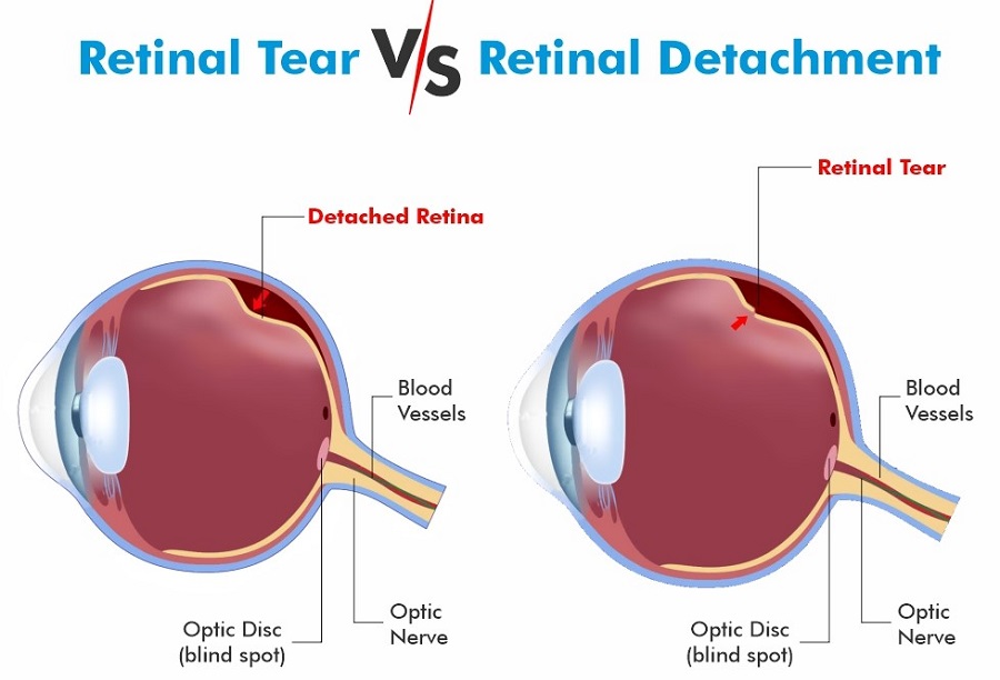
Retinal detachment is a serious condition that occurs when the retina is separated from the tissue to which it is attached, from the eye vessels that carry oxygen and nutrients to it.
If retinal tears are not recognized and treated, vitreous fluid may accumulate under the retina and cause retinal detachment.
Retinal detachment is a serious problem that can progress rapidly and lead to vision loss and should not be neglected.
What is Retinal Tear?
The retina is a thin tissue consisting of nerves that covers the inside of the eye. In a young and healthy eye, the retina is in direct contact with the vitreous fluid. With some diseases and aging, deterioration in the structure of this fluid occurs. These disruptions cause the retina to separate from the vitreous fluid. Retinal tears occur even if stronger shrinkage occurs at the points where the vitreous fluid is too attached to the retina.
What are The Causes and Types of Retinal Detachment?
Retinal detachment is not seen in the same way in all people. The types of this disorder, which causes serious problems in daily life, can be listed as follows;
-
Torn Retinal Detachment:
Retinal detachment tear and visual function problems are seen quite frequently compared to retracted and serous retina types.
The retina is surrounded by vitreous gel. This problem may occur as a result of the liquefaction of this gel and the separation of the posterior region of the viteus.
-
Retracted Retinal Detachment:
When this condition, which can occur in the vitreous or the upper parts of the retina, is seen, tearing is usually not seen.
However, in cases where the shrinkage is excessive, tears may also occur. Retracted retinal detachment occurs when the retina or inner membranes pull on the retina. In this case, the retina is pulled rather than torn.
Causes of retractive retinal detachment include;
- Diabetic retinopathy
- Formation of penetrating eye sores
- Premature retinopathy cases are included.
-
Exudative (Serous) Retinal Detachment:
Serous retinal detachment occurs when the retina is damaged. Serous retinal detachment is usually seen due to conditions such as inflammation or tumoral retina. It is not possible to see the tear in the retina, which has a flat surface.
The main causes of serous retinal detachment can be listed as follows;
- Posterior Scleritis
- Hypertension
- Harada Disease
- Pregnancy
- Tumors (Potential Leap)
What are The Symptoms of Retinal Detachment and Tearing?
Symptoms of retinal tear and retinal detachment develop rapidly and often appear out of nowhere. The reason for this is retinal detachment, a condition that develops suddenly and can progress rapidly. They require urgent intervention. If they are not intervened, they can result in separation and cause permanent vision loss.
Symptoms of retinal detachment and tearing are as follows:
- Seeing floating objects in the eye and gradually increasing the size of these objects
- Experiencing shadows in peripheral vision
- Blurring of vision and loss of vision
- Seeing a black curtain moving from one direction to the other in the field of vision
- Sudden flashes of light
- Such symptoms occur. If such symptoms are encountered, early intervention is important for treatment.
Situations with High Risk of Retinal Tear
-
Being High Myopic:
Being nearsighted above the number 3 increases the risk of retinal rupture 10 times. As the degree of myopia increases, the risk of tear formation also increases.
-
Retinal Thinning (Degeneration) Areas:
Retinal thinning areas are found in 8% of the normal population. In these thinning areas, tears may develop later.
-
Having Retinal Tear in the Other Eye:
8% of patients with a retinal tear in one eye also develop a retinal tear in the other eye.
-
Having a Family History of Retinal Tear:
Having a previous retinal tear in family members is one of the factors that increase the risk.
-
Considered Severe Traumas:
For example, sports injuries such as punching the eye, tennis ball, etc. or accidents that cause the eye to be punctured increase the risk of retinal tear.
-
Having Cataract Surgery:
After a successful cataract surgery, the risk of retinal tear is slightly increased. However, the risk of retinal tear increases 15 times after cataract surgeries with problems (posterior capsular rupture).
This shows the importance of performing cataract surgery in experienced hands.
How is Retinal Detachment and Tear Diagnosed?
The patient’s pupils are dilated with the help of drops, and retinal examination is performed with the help of a device called a biomicroscope. In this way, the hole, tear and detachment area in the retina are determined. Sometimes intraocular bleeding may also occur and make direct examination difficult. In such cases, an eye ultrasound may be required.
What are The Treatment Methods for Retinal Detachment and Tear?
Different treatment options can be applied to patients with retinal detachment and retinal detachment, depending on the condition of the disease. All of these are surgical operations that include different procedures. If there is only a tear in the retina instead of separation, laser suturing is performed.
The details of the relevant treatment methods are as follows:
-
Argon Laser:
It is a microsurgical application in which the tears are sutured with an argon laser. Patients are usually discharged the day after the surgery.
-
Vitrectomy:
Vitrectomy surgery is the most commonly used method in the treatment of retinal detachment today.
In the surgery, very small holes are made in the eye and the eye is entered. And with the help of devices that make up to 10,000 incisions per minute, the vitreous gel filling the eye is cleaned.
Necessary treatments can be applied to the retina as the pulling and tears are seen.
The detachment is removed by resorption of the fluid under the retina. At the end of the surgery, gas or silicone is injected into the eye.
Necessary laser treatments during surgery can be performed while in the eye.
-
Scleral Compression Technique:
Sometimes it is applied alone and sometimes in addition to virectomy. It is the placement of strips obtained from silicone under the conjunctiva. The strips are approximately 4 mm thick and this surgery is performed externally.
-
Pneumatic Retinopexy (Intraocular Tamponad Procedure):
In case of early detection of retinal detachment, it is applied as gas injection into the eye. After the procedure, argon laser is applied.
What Should Be Done After Retinal Detachment Surgery?
- In cases where gas or silicone oil is applied into the eye, the patient may need to lie/sit in the prone position for the period specified by the surgeon (it can vary from 1 day to 2 weeks).
- Just like after cataract surgery, the patient uses drops gradually for a month.
- The first week is a particularly important time to prevent the eye from getting infected. It is very important not to be in very crowded places, dusty, dirty environments, not to touch or rub the eyes, and to keep them clean.
- Generally, it is ideal to take a week off and rest in the first week after the surgery. This period may vary depending on the severity of the disease.
- When air/gas is left in the eye, it is prohibited to travel to high altitudes such as mountains or to travel by plane. The surgeon must be consulted.
Again, the first month after the operation;
- Heavy sport,
- Weight lifting
It is beneficial to avoid strenuous activities such as frequent bending, straining, and severe coughing.
What are The Complications of Retinal Tear?
- Retinal tear can lead to serious complications when left untreated or unsuccessful.
- Due to a poorly treated tear, the retinal layer can be completely separated. This condition is called retinal detachment.
- It is a condition that requires immediate medical attention. If the retina cannot be repositioned, vision loss can occur.
- If a tear or hole occurs in the area close to the center of the retina, that is, in the macula area, it can have serious effects on vision. And permanent loss of central vision may develop.
- Sometimes bleeding can develop after a retinal tear. It can seep under the retina.
- Although not common, infection or inflammation can occur. Eye pain, redness, and vision changes may occur. In some cases, an increase in intraocular pressure may develop.
What Complications Cause Retinal Tear?
A retinal tear can also cause other conditions.
When treated, positive results are obtained.
However, different eye diseases may occur if treatment is delayed, untreated, or not treated successfully.
-
Retinal Detachment:
When the tear is not treated, it is called the separation of the retinal layer.
-
Macular Tear:
It is a tear in the center of the retina.
-
Subretinal Neovascularization:
It is the formation of new blood vessels and leakage of fluid under the retina after a retinal tear.
-
Epiretinal Membrane:
It is the formation of membranes that adhere to the retina in the eye after a tear.
-
Cataract:
It is the clouding of the natural lens of the eye.