What is Otosclerosis (Inner Ear Calcification)?
Otosclerosis (Inner Ear Calcification)
Hearing occurs as a result of sound waves reaching the eardrum and vibrating the eardrum, this vibration is transferred to the inner ear fluids with the movement of the hammer, anvil and stirrup bones in the middle ear, converted into electrical energy in the nerve endings in the inner ear, and reaches the auditory center in the brain via the auditory nerve.
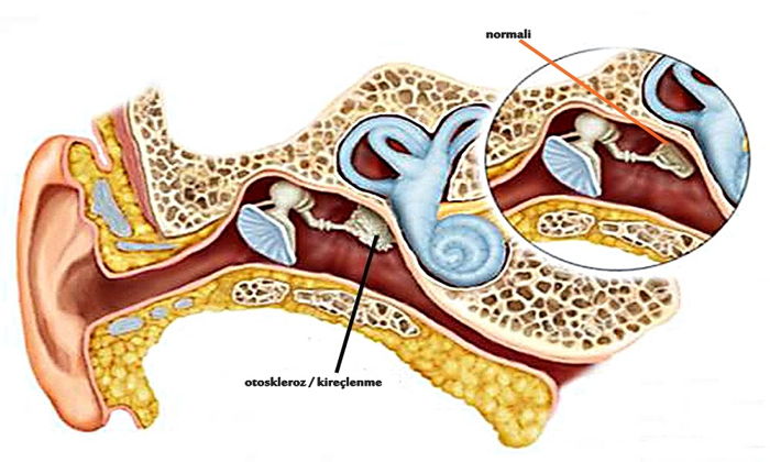
Otosclerosis is defined as calcification of the wall parts of the stirrup that is adjacent to the inner ear, and the restriction of the movements of the stirrup ossicles. Immobilized ossicles cannot transmit sound waves and hearing loss occurs. At first, the inner ear is intact and only sounds cannot be transmitted. However, in the future, calcification may also affect the inner ear wall and cause neural-type hearing loss.
How Does Hearing Occur?
Our ears are the organs on both sides of our head that are responsible for transforming the vibrations, sound waves, produced by certain substances circulating in the air, into messages that our brain can process. The clear transmission of sounds to the brain and their good perception indicate the quality of hearing, that is, the ears are working well.
The ear has three main parts, the outer ear, middle ear, and inner ear. And these 3 main sections have different functions from each other. The outer ear collects sound waves, ie vibrations, in the external environment and transmits them to the middle ear.
The middle ear is responsible for transmitting sound vibrations from the eardrum to the inner ear fluids by mechanical pressure. The inner ear undertakes the task of converting these pressure waves transmitted from the middle ear into messages that our brain can perceive.
The 3 bones in the middle ear are as follows:
- Hammer (Malleus): It is the outermost bone attached to the eardrum.
- Anvil (Incus): It is the bone in the middle of two bones and is responsible for transmission.
- Stapes: It is the bone in charge of transmitting vibrations transmitted from the anvil to the fluids in the inner ear. Sound vibrations transmitted by the stirrup (stapes) to the oval window and inner ear fluids cause fluctuations in the inner ear fluids.
These fluctuations are transformed into electrical signals, that is, messages that the brain can perceive, by the sensory organs in the structure of the inner ear called the cochlea, which is the auditory part of the inner ear. These signals are transmitted to the brain stem, which is connected to the auditory nerve, and from there to the brain. In this way, hearing takes place.
What are the Types of Hearing Loss?
After examinations called audiometric tests, hearing loss is divided into 5 types.
These:
- Conductive Hearing Loss: It is observed as a weaker hearing due to any pathology in the sound conduction path. If the patient has pathology/rupture in the middle ear and/or eardrum, the sound is perceived weaker in Eustachian tube pathologies.
- Neural (Sensorineural) Hearing Loss: It is the loss that occurs as a result of damage to the auditory nerves in the inner ear.
- Mixed (Mixed) Type Hearing Loss: The presence of sensorineural hearing loss in addition to conductive hearing loss is called mixed hearing loss. Generally, both ears are affected by this disease in patients, but the level of involvement of these two ears may not always be the same. In fact, it is generally seen that one ear is more affected than the other.
- Functional hearing loss: Losses that do not have an organic pathology to explain the underlying hearing loss. It is generally considered to be emotional hearing loss. This condition is called pseudohypacusia.
- Central Hearing Loss: It develops as a result of damage to the hearing center in the brain.
What Causes Otosclerosis?
The cause of otosclerosis is still not clearly defined. However, there is a hereditary (familial) transmission characteristic of the disease. For example, in some cases such as the presence of measles virus and pregnancy, worsening of the course of otosclerosis may be observed. In some cases such as these, otosclerosis may have such links. It is recommended to get information from your doctor about these conditions. If this disease is left untreated, hearing loss increases until late middle age. Therefore, otosclerosis is defined as a progressive disease. Although the disease is most common in middle-aged women, it can also be seen in men and children.
What are Otosclerosis (Inner Ear Calcification) Symptoms?
The most common symptom of inner ear calcification, or otosclerosis, is hearing loss. Hearing loss that occurs in otosclerosis disease occurs as the inability to hear low-level or high-pitched sounds. Hearing loss can progress over time. In case of otosclerosis, hearing loss is mostly experienced bilaterally. In addition to symptoms such as hearing loss, ear pain,
Otosclerosis symptoms can be listed as follows;
- The inner ear structure that provides hearing is also the center of balance. Otosclerosis, ie inner ear calcification symptom, can also occur as dizziness and balance problems.
- Tinnitus can be due to different reasons, but it can also be experienced as a symptom of otosclerosis. At the same time, ear pain can be seen among the symptoms of otosclerosis.
- One of the symptoms of otosclerosis is speaking in a low voice. In the presence of otosclerosis, the person hears his own voice louder and therefore can speak in a low voice.
- Otosclerosis patients hear less ambient noise in noisy environments and more clearly the speech of people. This condition, known as Willis paracusis, is typical in patients with otosclerosis.
How is Otosclerosis (Inner Ear Calcification) Diagnosed?
Otosclerosis (inner ear calcification) is diagnosed by an ear, nose and throat specialist doctor. The patient’s history is important during the examination. Ear examination is usually normal. Sometimes a slight reddish reflex can be observed behind the eardrum.
This indicates that the disease is in the initial stage. Your ear is being examined to rule out ailments that have symptoms similar to those of otosclerosis. Hearing tests are performed to determine the degree of hearing loss experienced.
- In the audiogram hearing test for the diagnosis of otosclerosis, the lowest sounds heard at different pitches and frequencies are determined.
- With the tympanometry test, it can be revealed whether there is an calcification in the ossicular system by evaluating the flexibility of the eardrum.
- With the acoustic reflex test, the effect of this calcification on the stirrup is clearly evaluated.
- In some cases, Computed Tomography (CT) imaging may also be requested in order to better see the condition of the bones and tissues in the ear and to clarify the diagnosis.
What is a Tuning fork Audiogram (Otosclerosis Audiogram)?
The definitive diagnosis can only be made during the operation in patients who have conductive hearing loss despite all diagnostic tests and tests performed before the patient is treated with surgery. When the middle ear is checked during the surgery in some patients who are operated because of the diagnosis of otosclerosis, it can be understood that the cause of hearing loss is other than otosclerosis. This is why the definitive diagnosis can be made during surgery in patients with conductive hearing loss. In these cases, it becomes more difficult to correct this hearing loss with surgery, and sometimes even it cannot be corrected depending on the cause of the hearing loss.
What are Otosclerosis (Inner Ear Calcification) Treatment Methods?
If otosclerosis is at the initial stage, the patient can be followed up without any treatment. In this process, the hearing status of the patient should be followed closely by performing a hearing test at regular intervals. However, since otosclerosis is a disease that can progress over time, it should be treated sensitively.
Otosclerosis treatment is generally carried out as follows;
- Hearing aids can be used in the treatment of otosclerosis. However, hearing aids do not cure otosclerosis, they only eliminate the problems caused by the hearing loss.
- Otosclerosis surgery: If the patient’s hearing loss is advanced and the disease is predicted to progress in the examinations, a surgical procedure can be performed. In the surgery, some or all of the problematic bone is removed and an implant that will provide hearing is placed in its place.
- Fluoride therapy can be used in some patients. Fluoride treatment is mostly preferred in some cases for a temporary period before preparation for surgery.
How is Otosclerosis (Inner Ear Calcification) Surgery Performed?
The surgery to be applied in otosclerosis disease is stapes surgery. The procedure is usually performed under general anesthesia. A small incision made inside the ear canal is followed and the middle ear is entered behind the eardrum. Malleus, incus and stapes (hammer, anvil and stirrup) connections and mobility in the middle ear are evaluated.
The presence of mobility of the malleus and incus and the absence or severe loss of the mobility of the stapes bone make the definitive diagnosis. After this point, the operation is terminated by replacing the stapes bone with a Teflon prosthesis.
After the operation, the patient is discharged after being followed up in the hospital for 1-2 days. One week later, the external auditory canal of the patient who comes to the control is cleaned and the follow-up continues. The patient’s hearing is evaluated by performing control audiometry tests in the 1st month and 3rd month postoperatively.
What are the Risks of Otosclerosis (Inner Ear Calcification) Surgery?
The risks of otosclerosis surgery can be listed as follows;
- Sometimes, the hearing problem may not be corrected with surgery. The patient may not benefit from the surgery. This is a very rare condition.
- Although it is very rare, the hearing of patients may be adversely affected after otosclerosis surgery.
- Bleeding, wound new infection, middle ear infection can be experienced as general surgical complications.
- The facial nerve normally passes close to the stirrup where the prosthesis will be attached. Normally, there is a bony structure on the facial nerve that protects the nerve. In some patients, this bone may not be found anatomically. The facial nerve can pass openly. It is not possible to determine this condition preoperatively. Or the facial nerve may rest on the stirrup. In such cases, surgery may not be performed to prevent facial paralysis by damaging the nerve.
- Patients may experience hearing loss again due to calcification or slippage of the implant in the long term.
However, these complications are very rare. Most of the patients get rid of the complaints they experience after the surgery. It is very important that the surgery is performed in fully equipped centers by experienced teams.
What Should Be Considered After Otosclerosis (Inner Ear Calcification) Surgery?
It is important for patients to follow some rules in the first 2-3 months after otosclerosis surgery.
Things to consider after otosclerosis surgery can be listed as follows;
- It is necessary to stay away from daily life activities that will increase intracranial pressure after surgery. Heavy sports can damage the implant used in surgery by increasing intracranial pressure from sports such as lifting weights. However, light cardiac activities such as walking can be done easily.
- The ear should be protected against impacts.
- It is necessary to be protected from sudden pressure changes. Situations where the pressure in the cabin suddenly changes during airplane journeys can cause problems for patients. Before boarding the plane, patients can be given medication against these pressure changes. Likewise, diving under water is among the activities that should be avoided in the postoperative period, as it can create a pressure difference.
- After the healing of the wound in the external ear canal (2-3 weeks), the patient can easily enter the sea or the pool.
These precautions are important for the first 2-3 months after surgery. Afterwards, patients can continue their normal routine lives.