Treatment Options For Cartilage Injuries
Articular cartilage is a very special structure designed to carry loads and provide painless movement in the joint for many years. It serves as a cushion by covering the faces of the bones that make up the joint facing each other. Articular cartilage can be damaged in several ways. Over the years, it gets old, softens first, then fringes out, and the bone beneath it emerges. Popularly known as” calcification”, this condition is called osteoarthritis or arthrosis.
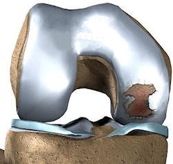
It is the result of wear and obsolescence that occurs with age. This widespread abrasion that occurs is irreversible and requires medication first and then surgical treatments. There is no known treatment method for osteoarthritis that regenerates cartilage today. However, in young individuals, regional damage to the articular cartilage may occur due to bumps, especially during sports. In such cases, only part of the joint cartilage is damaged and the rest is intact, so cartilage regenerating treatments can be done.
The topic of this article is the treatment options for regional cartilage injuries in these young individuals.
In adults, the healing ability of articular cartilage is virtually nonexistent. Unlike other tissues in the body, articular cartilage does not regenerate itself after injury. Surgical interventions are absolutely necessary to create the healing response in cartilage.
What are the symptoms of cartilage injuries?
The most important symptom of cartilage injuries is pain that focuses on the relevant area of the joint. Joint swelling may occur due to increased fluid in the joint. This swelling increases with activity or sport, decreases with rest. Joint may cause complaints such as tripping, jamming, locking. If there is a broken piece, patients may feel a piece that travels freely within the joint. This piece is called a joint mouse. In cartilage injuries that are above a certain size, arthrosis may develop over time as the load bearing properties of the joint will be impaired.
How do cartilage injuries occur?
Cartilage injuries occur most often as a result of blows from insertion. This can occur due to bumps from direct insertion during a sporting event or after a traffic accident. It is most commonly seen in the knee joint. Cartilage injuries in the knee may be accompanied by meniscus and cruciate ligament injuries, and their symptoms may also occur.
In addition, regional cartilage injuries may also occur in fractures or dislocations involving the joint. Kneecap bone (patella) and shoulder dislocations are the most common examples. Another form of formation is cartilage damage that occurs as a result of the disease called “osteochondritis dissecans”, which occurs due to the eating disorder of the bone below the articular cartilage and where this non-feeding area is separated from the intact bone and becomes free.
How to diagnose cartilage injuries?
For the diagnosis of cartilage injuries, your doctor first listens to the pattern and symptoms of the event. If he thinks there may be cartilage damage with a detailed examination he will then resort to imaging methods. X-rays are taken first. If you have a fracture or osteochondritis dissecans, you can be diagnosed here. If there are dislocations related to the joint, the symptoms of this can be determined.
Magnetic resonance imaging (MRI) of the joint can then be performed if your doctor deems it necessary. Diagnosis of large and full coat cartilage damage can be easily verified by MRI. But standard MRI may not always be successful in diagnosing cartilage injuries that are not full-fold. In some cases, your doctor may ask for an MRI-arthrographic examination, which is performed by placing contrast agents in the vein or into the joint.
If other symptoms strongly suggest cartilage injury, your doctor may recommend arthroscopy for both diagnostic and therapeutic purposes. The diagnosis of cartilage damage can be made with certainty by arthroscopy.
What are the treatment options for cartilage injuries?
Cartilage damage in the non-load-bearing area of the joint and less than 1 cm2 can only be followed by intermittent follow-up if it does not lead to complaints. However, treatment is necessary for cartilage injuries greater than 1 cm, which are located in the region that gives symptoms, carrying loads. Drugs, such as glucosamine supplements, physical therapy methods and injections of hyaluronic acid into the joint can be done.
However, they do not have therapeutic properties and only suppress the symptoms for a period of time.
In young adults, the treatment of cartilage injuries is surgery. Treatment often begins with arthroscopy and they are also corrected if there are other accompanying problems within the joint. The procedure for cartilage can then be performed with arthroscopic or open surgery.
Microfracture method
It is a method applied in cartilage injuries that are limited and smaller than 3cm2. After the damaged area is cleared of cartilage residues, holes extending to the bone at 5 mm intervals and a few mm deep are opened. The stem cells in the bone marrow are used to treat the disease.
Stem cells, which are localized into the blood clot that forms at the site of damage, have the ability to develop into cartilage-like cells when the appropriate environment is provided.
In recent years, roof implants called Matrix have been developed to better hold and organize this clot into the damaged area.
These membrane-shaped tissues, made from most collagen, can be affixed to the damaged area after micro-fracture. It is necessary to protect the joint from loading by using crutches for six to eight weeks after surgery until new cartilage-like tissue is formed.
Arthroscopic micro-fracture technique
The advantages of micro-fracture method are that it is an easy and inexpensive technique for the patient and the physician, it requires a single operation and it can be done with arthroscopy. It is often the first preferred method for small-scale cartilage damage. However, the most important disadvantage of the technique is that the repair tissue that is formed is “cartilage-like”. This tissue differs from normal cartilage and cannot be expected to function like normal cartilage for many years.
Mozaikplasty
It is the removal of cylindrical pieces of cartilage and bone 6-8 mm in diameter and 15 mm in length from the non-load-bearing region of the joint, and their transfer to the damaged area in the load-bearing region.
This technique is applied in damages below te 4cm2. It is most commonly applied in the knee and ankle joints. It can be done by arthroscopic or open method. The most important advantage is that a tissue is transplanted into the damaged area in the architectural structure of normal cartilage.
The main drawback is that normal cartilage in another part of the joint is sacrificed so that a limited number of tissue transplants can be performed and transplanted to the damaged area. In very large damages, tissue may also need to be removed from the opposite knee joint, which is sometimes intact.
Due to the characteristic of the technique, only 70% of the damaged area can be filled with transplanted cartilage, and the area between the transplanted cylinders is healed with a cartilage-like repair tissue. This technique is often more successful in minor cartilage damage. In areas of major injury, the normal shape of the joint can be difficult to establish. The post-operative period is similar to the micro-fracture method.
Cartilage transplantation
In recent years, the most researched and scientific developments on the field is cartilage transplantation. In this technique, a few millimeters of cartilage tissue is removed in the form of chipping from the non-load-bearing area of the joint when damage to cartilage is detected by arthroscopy first. This tissue is processed in a laboratory environment under sterile conditions and produced by replicating the cartilage cells in it.
After a few weeks of this procedure, new cartilage cells are transplanted into the damaged area, this time with open surgery. In cartilage transplantation techniques called first generation, these cells were injected under a membrane-shaped tissue taken from tissues around the knee and stitched into the damaged area.
First generation cartilage transplantation
A tissue sample is taken, produced and injected under a cover-shaped cover in the damaged area.
The most important advantage of cartilage transplantation techniques is that the patient’s own cells can be transferred to the damaged area in the desired amount without damaging any tissue.
There is no size limit and tissue can be produced at the desired diameter and height. The resulting new cartilage tissue is much closer to normal articular cartilage.
The drawbacks of the technique are that it requires two surgeries and is expensive. Although it is a very new method, successful results have been reported in 80-90% of patients in follow-up over ten years in the world. The post-operative period is similar to other techniques.


