What is Skin Cancer?
Skin is one of the largest organs in our body. Skin cancer, also known as skin cancer, is a health problem that occurs as a result of the uncontrolled proliferation of melanocyte cells, which produce color pigments called melanin, which gives the skin its color. This condition, also called melanoma, can sometimes appear suddenly on the skin without showing any symptoms, and sometimes it can appear on a “mole” tissue that the person previously had.
As a result of the unusual change and proliferation of the structures of melanocyte cells, these cells have the potential to spread to other organs. Melanomas are less common than other skin cancers. Despite that; Approximately 3/4 of people who die due to skin cancer are affected by the deterioration of the structure of melanomas. Additionally, skin cancer is a disease that spreads extremely quickly. Therefore, just like other types of cancer, early diagnosis is extremely important in treating skin cancer.
What are The Factors That Cause Skin Cancer?
-
Exposure to Ultraviolet (UV) Ray:
UV rays are one of the main risk factors for melanoma skin cancer. It damages the DNA of skin cells and skin cancer begins. Sunlight is the main source of ultraviolet rays. It is said that solarium is another source of UV rays. People who are exposed to excessive UV rays from these sources increase the risk of many skin cancers, including melanoma.
-
UVA Rays:
It causes cell aging and can damage cell DNA. It is thought to cause long-term damage to the skin, such as wrinkles, and to play a role in the development of some skin cancers.
-
UVB Rays:
It is the main ray that causes sunburns and can directly damage cell DNA. It is thought to cause most skin cancers.
-
UVC Rays:
It cannot pass through the atmosphere. Therefore, it is not found in sunlight. For this reason, it does not cause skin cancer.
-
Solarium:
Research has shown that the risk of melanoma skin cancer is higher in people who frequently go to solariums to tan. UV lamps used for tanning in solariums must be “ultraviolet lamps” and are labeled with the phrase “Continuous exposure to UV rays can cause premature aging of the skin and skin cancer.” should be.
Additionally, it is thought that placing a label stating “regular medical check-up is necessary for skin cancer” may be a warning for users who are constantly exposed to these rays.
Thus, the use of ultraviolet products (black light lamp, mercury vapor lamp, high pressure xenon and xenon mercury arc lamp, plasma torches and arc sources, etc.), especially for young people/children under the age of 18 who are at risk of skin cancer and people with skin cancer in their family. It is aimed to reduce the counter tendency.
-
Moles:
Moles on our body are benign tumors and occur not only at birth but also during childhood and adolescence. Most moles never cause problems. However, people with many moles have a higher risk of developing melanoma.
-
Normal Moles:
It usually appears as a brown, tan or black spot on the skin. It can be flat or high and raised, round or oval, and is usually smaller than 6 mm.
Moles may occur at birth or during childhood and adolescence. New moles that appear on the body during adulthood should be checked by a doctor against possible skin cancer. When a mole develops on the body, it will remain the same size, shape and color for years.
Some moles will disappear on their own over time. Many people have moles, and most of these moles are harmless.
-
Dysplastic Nevus:
Dysplastic nevi (nevi is the plural form of nevus), or unusual nevi, often look at least slightly like normal moles, but have some features of melanoma. They are usually larger than other moles, have an unusual shape or color, and most do not become cancerous.
-
Congenital Melanocytic Nevus:
Moles present at birth are called congenital melanocytic nevi. It is estimated that the risk of developing melanoma in such moles that are present at birth is between 0-10%, depending on the size of the nevus.
People with large congenital melanocytic nevus have a higher risk of developing melanoma. For example; If the congenital nevi are smaller than the palm, the risk of melanoma is lower. On the contrary, the risk of melanoma increases significantly in congenital nevi of large size on the back or buttocks.
-
Fair Skin, Freckles and Light Hair:
The risk of melanoma in people with white skin and light hair is 10 times higher than in people with black skin.
The risk of skin cancer increases in people with red and blonde hair, white skin, blue or green eyes, or freckles.
-
Age:
Although melanoma is frequently seen in young people between the ages of 15-29, it is most common in the 25-29 age group (especially in young women). However, it can also be seen in older ages.
-
Gender:
Considering their frequency and biological differences, skin cancers are divided into two groups: malignant melanoma and non-melanoma skin cancers. While it is the 4th most common cancer type in young adult men, that is, men between the ages of 25-34, it is the most common cancer type in women after breast and gynecological cancers.
-
Dealing with Welding and Metal Work:
It has been shown to increase the risk of melanoma in the eyes.
-
Phototherapy (Light Therapy):
UV rays exposed to people with certain skin diseases, such as psoriasis, due to treatment, increase the risk of squamous cell skin cancer.
What are The Symptoms of Skin Cancer?
The most important symptom of skin cancer is the formation of a new spot on the skin or a change in the size, shape or color of the spot.
Another important sign is that the skin scar looks different from other scars on your skin.
Abnormal growth and shape change of moles, lesions, skin peeling, swollen, crusty and bleeding wounds are common symptoms of skin cancer.
Here are some changes to look out for when checking your skin for any signs of skin cancer:
- Changes in the size, shape or color of moles
- Itchy and painful moles
- Asymmetrical moles with irregular edges
- Bleeding and non-healing wounds
- Red or skin-coloured shiny bumps on the skin
- Newly formed or discolored moles and lesions
- Swelling under the skin in areas such as neck, armpit or groin
- Feeling tired
- Lesions that grow and swell in the form of lumps
Follow-up of moles is important for early diagnosis of melanoma. A method called the “ABCDE rule” is used to easily detect differentiation in the follow-up of moles.
-
A (Asymmetry):
One half of the mole resembles the other half in color and/or shape,
-
B (Border Irregularity):
The borders of the mole are indented and irregular,
-
C (Color):
The color of the mole is not homogeneous (if two or more colors such as brown, black, red, gray and white are present together or if there is a variegated appearance),
-
D (Diameter):
If the mole is larger than 6 mm in diameter, that is, roughly the diameter of a pencil with eraser,
-
E (Elevation):
This is because there is swelling on the mole.
If the person observes these conditions, he/she should consult a doctor.
What are The Types of Skin Cancer?
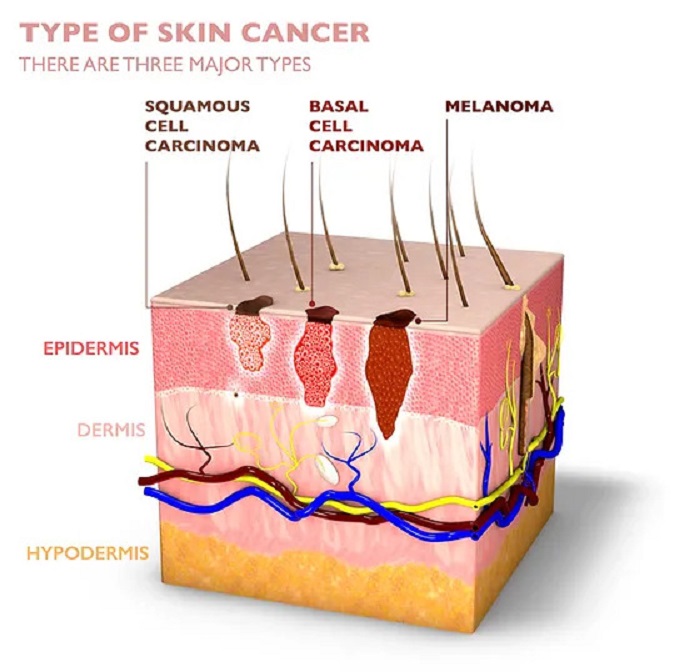
- Basal cell carcinoma
- Squamous cell carcinoma
- Melanoma
- Kaposi’s sarcoma
- Merkel cell carcinoma
- Sebaceous gland carcinoma
What are The Signs and Symptoms of Skin Cancer Types?
Type of Basal Cell Carcinoma Skin Cancer Symptoms:
Basal cell carcinoma is the most common skin cancer.
It most often occurs on sun-exposed areas of the body, such as the neck or face.
Symptoms of basal cell carcinoma type skin cancer may appear as described below:
- Pearl-like, mother-of-pearl or waxy swelling
- Flat, flesh-colored or brown scar-like lesion
Squamous Cell Carcinoma Type Skin Cancer Symptoms
Squamous cell carcinoma occurs most often on sun-exposed areas of your body, such as the face, ears, and hands. In people with darker skin tone, squamous cell carcinoma is a type of skin cancer that is more common to develop in skin areas that are not frequently exposed to the sun.
Squamous cell carcinoma type skin cancer may appear as described below:
- Hard, red nodule
- Flat lesion with scaly or crusty surface
Melanoma Type Skin Cancer Symptoms
Melanoma can occur anywhere on your body, on skin with no other symptoms or in a skin mole that turns into cancer.
It is most commonly seen on the face or trunk of affected men.
In women, this type of cancer occurs most often in the calves. Melanoma can occur on skin that has not been exposed to the sun in both men and women.
Melanoma can occur in all people, regardless of skin color. In people with darker skin tone, melanoma tends to appear on the palms or soles of the feet or under the fingernails and toenails.
Signs of Melanoma Type Skin Cancer İnclude:
- Large brownish spot with dark spots.
- Skin mole that changes color, size, or sensation, or where bleeding is observed.
- Small lesion with irregular borders and areas that appear red, white, blue or blue-black.
- Dark lesions on the palms of the hands, soles of the feet, fingertips or toes, or on the mucous membranes lining the mouth, nose, vagina or anüs.
Symptoms and Signs of Less Common Skin Cancers:
Other Less Common Types of Skin Cancer İnclude:
Kaposi Sarcoma Type Skin Cancer:
This rare type of skin cancer occurs in the blood vessels of the skin and causes red or purple patches on the skin or mucous membranes.
Kaposi’s sarcoma usually occurs in people with weakened immune systems, such as people taking drugs that suppress the natural immune system, such as AIDS patients and organ transplant patients.
Other people at increased risk of Kaposi’s sarcoma include young men living in Africa or older men of Italian or eastern European Jewish heritage.
Merkel Cell Carcinoma:
Merkel cell carcinoma causes hard, shiny nodules that occur on or just under the skin and in hair follicles. And merkel cell carcinoma is most often found in the head, neck and trunk.
Sebaceous Gland Carcinoma:
This rare and aggressive cancer originates from the oil glands in the skin. Most often appearing as a hard, painless nodule, sebaceous gland carcinoma can occur anywhere, but most often occurs on the eyelid and is often confused with other eyelid problems.
How Many Stages Does Skin Cancer Consist of?
Skin cancer is evaluated by dividing it into stages. There are 5 stages of skin cancer in total and treatment & prognosis are determined according to the stages.
-
Skin Cancer Stage 0 (Carcinoma in Situ):
Stage 0 is the stage where the cancer is still limited to the skin surface and has not yet spread to surrounding tissues. At this stage, cancer can be completely cured.
-
Skin Cancer Stage 1:
Stage I is the stage where cancer has spread through slightly thickened layers of skin but has not spread to surrounding tissues. At this stage, cancer can be treated.
-
Skin Cancer Stage 2:
Stage II is the stage when cancer has spread through the deeper skin layers and spread to surrounding tissues. At this stage, cancer may be more difficult to treat.
-
Skin Cancer Stage 3:
Stage III is the stage when cancer has spread to deep tissues and spread to nearby lymph nodes. At this stage, treating cancer may be more complex.
-
Skin Cancer Stage 4:
Stage IV is the stage when cancer has spread to other parts of the body. Treating cancer at this stage can be more difficult and often only focuses on controlling symptoms.
How is Skin Cancer Diagnosed?
Diagnosing skin cancer involves detecting abnormalities on the skin and determining the type of cancer. Early diagnosis is critical to successfully treating skin cancer.
-
Dermatoscopic Examination:
Dermatoscopic examination is an imaging method that allows in-depth examination of lesions and moles on the skin. Dermatologists use a special instrument called a dermoscope to magnify the skin surface and identify abnormalities.
-
Biopsy:
Tissue samples are taken from suspicious lesions on the skin and a biopsy is performed. The biopsy result helps determine the type and stage of cancer.
-
Histopathological Examination:
Tissue samples taken during biopsy are examined under a microscope by pathologists. This examination helps determine the presence and spread of cancer cells.
-
Display Methods:
In some cases, imaging methods can be used to determine whether skin cancer has spread to other organs. The spread of cancer can be evaluated using methods such as MRI, CT scans and PET scans.
Treatment Methods for Skin Cancer
Treatment for Small Fragment Cancer Cells
Curettage and Electrodessication (C and E):
It is a form of treatment used for squamous cell types of cancer. The procedure involves removing the skin cancerous surface with a scraping tool (curette) and cutting its base with an electric needle.
-
Laser Treatment:
It is a treatment method used to vaporize skin lesions with an intense beam of light.
Laser treatment prevents lesion growth and causes minimal damage to the part of the lesion; It reduces the risk of symptoms such as bleeding and swelling.
-
Cryosurgery:
It can be used in the treatment of actinic keratosis and some small skin cancers. Liquid nitrogen is used to destroy abnormal cells by freezing. After the diseased area dissolves, dead tissues separate and fall off. The procedure may need to be repeated to completely destroy the growing tissue. Cryosurgery is generally painless; However, pain and swelling may occur at the wound site after the procedure. A white scar may occur in the treated area.
-
Photodynamic Therapy:
Treatment of superficial skin cancers; It is a form of treatment that combines photosensitizing drugs and light. Cell sensitivity is achieved with a liquid drug, and a light that can destroy cancer cells is shined on this area.
Treatment for Larger Cancer Cells
-
Simple Excision:
The cancerous tissue and the surrounding healthy skin lesion are excised. In cases of necessity (wide excision), removal of the normal skin surrounding the tumor may also be recommended.
-
Mohs Surgery:
During this surgery, your doctor removes the cancerous part layer by layer and continues the process until no abnormal cells remain.
After examining each layer under a microscope, it is ensured that all growth is removed and the process is completed.
-
Radiation Therapy:
It is the process of neutralizing cancer cells using high-energy rays (X-rays and protons). Radiation therapy is sometimes used after surgery in cases where there is an increased risk of the cancer returning. It can also be considered an alternative method for people who cannot undergo surgery.
Treatments for Cancers That Have Spread Beyond the Skin
-
Chemotherapy:
It is the process of killing cancer cells using strong drugs. However, if the carcinomas have reached the lymph nodes or other parts of the body, it can be used together with treatments such as radiation and drug therapy, as well as chemotherapy.
-
Targeted Drug Therapy:
It is a treatment method combined with chemotherapy that weakens the target and causes cancer cells to die.
-
Immunotherapy:
It is a form of therapy that enables your immune system to fight cancer. However, your body’s disease-fighting immune system may not be able to resist cancer because cancer cells produce proteins that blind the immune system cells. Immunotherapy interferes with this process, although immunotherapy may be considered when the cancer is advanced and cannot be addressed by other options.
What are The Ways to Prevent Skin Cancer?
There are risk factors that cause skin cancer and some conditions that need to be applied to protect against UV rays.
Things to do to protect from skin cancer are as follows:
-
Protect Your Skin:
If you are not sunbathing, put on some clothes, wear a wide-brimmed hat and protect your skin as much as possible. You can protect your eyes by wearing sunglasses that block at least 99% of UV rays.
-
Try to Sit in the Shade:
Do not sunbathe between 10:00 and 16:00, when the sun’s rays are the harshest. Limit your exposure to direct sunlight to the periods specified by experts.
-
Do not tan in the solarium:
Tanning in a solarium can contribute to skin cancer and cause long-term damage to your skin.
-
Pay Attention to The Expiration Date of Cosmetic Products:
Use broad-spectrum sunscreens with a sun protection factor of at least 30. Be sure to apply sunscreen every 2 hours and after swimming and sweating.


