


Tinnitus
Tinnitus in medicine; It is a condition that can seriously reduce the quality of life of people. The causes of tinnitus are described as sizzling, buzzing, hearing noises like water noise. Almost 10% of people experience tinnitus at some point in their lives.
Tinnitus may be intermittent or continuous. The timbre of sound can be thick or very thin, affecting one ear or both. Tinnitus can make this person very uncomfortable when it is continuous.
This discomfort can affect people’s normal lives to the extent that can go up.
Tinnitus is mainly divided into objective and subjective.
Objective tinnitus; this is the case where not only the sick person but others also hear these sounds.


Subjective tinnitus; is the perception of sounds only by the patient.
What Are The Causes of Tinnitus?
There are many possible causes of tinnitus. These reasons can often be simple and benign, as well as more serious problems.
Common causes are;
- Damage to the auditory nerve endings in the inner ear
- Ear wax
- Problems in the eardrum
- Ear infections
- Allergic rhinitis
- Sinusitis
- Trauma to the ear area
- Accumulation of fluid in the middle ear
- Hardening of the joints of the middle ear bones
- Contraction of the muscles in the middle ear,
- Expansion of the Eustachian tube (maybe due to pregnancy or weight loss)
- Arterial anomalies in the middle ear
Continue
- Vein enlargement in head and neck
- Tumour in the nerves that provide balance and hearing (acoustic neurinoma)
- Allergy
- Low or high blood pressure
- Blows to the head and neck
- Neck calcification
- Some medications (rheumatism medications, antibiotics, aspirin, etc.)
- Diabetes
- Thyroid diseases
One of the most common causes of tinnitus is; damage to the auditory nerves of the inner ear. This occurs especially at an advanced age.
Tinnitus Treatment
There is usually no special treatment in cases of tinnitus. To treat tinnitus, first attempt to identify underlying treatable conditions that may be associated with your symptoms. If tinnitus is caused by a health condition, your doctor may take steps to reduce noise. Some X-rays and balance tests may be needed. However, the cause of tinnitus may not be found despite all tests.
There are also some medications used in the treatment of tinnitus. Although these drugs provide relief, they may not be sufficient for treatment. Most people get used to the tinnitus and learn to live with it. Ignoring instead of standing on it can also provide relief in tinnitus.
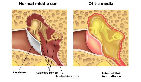
Middle Ear Inflammation
Middle ear; hammer, anvil and stirrup, which contain small bone structures, separated from the eardrum and the outer ear canal, called the eustachian tube with a narrow opening behind the nose and throat. The eustachian tube is a channel found in the human body in pairs. It plays an important role in regulating air pressure in the middle ear, regenerating air and clearing secretions. Another related structure that may play a role in middle ear inflammation is adenoids.
Adenoids, located in the upper part of the back of the nose, which is thought to play an important role in the activity of the immune system in the body is a double structure. Eustachian tubes have an important place for the health of the middle ear because they are located near the nasal and throat opening and may cause blockage in the eustachian tube in case of infection.

Causes Middle Ear Inflammation?
Inflammation of the middle ear is a disease caused by bacteria or viruses. Infection is usually; colds, flu, allergies, such as nasal canals, throat and eustachian tubes are caused by swelling of the disease.
What Are The Symptoms of MiddleEear İnflammation?
The incidence of middle ear inflammation increases in autumn and winter when upper respiratory tract infections are more common. Although the disease can be seen at any age, it is more common in childhood and it is known that more than 80% of children under 3 years of age experience at least one otitis media.
Symptoms of Middle Ear Inflammation in Children:
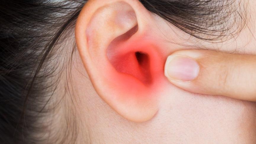
- Ear pain (especially in supine position)
- Difficulty sleeping
- Crying more than usual
- Don’t be more restless than usual
- Difficulty in hearing or difficulty responding to environmental audible stimuli
- Loss of balance
- Fire
- Ear discharge
- Headache
- Anorexia
Symptoms of Middle Ear Inflammation in Adults:
- Earache
- Ear discharge
- It is manifested by hearing loss.

How İs The Diagnosis of Otitis Media Diagnosed?
Diagnosis of middle ear inflammation is often made by the patient’s history and physical examination of the physician. In the physical examination of the ear, light instruments called otoscopes are used to see the structures within the ear. Diagnosis is made when the physician detects fluid accumulation in the middle ear and signs of infection.
How to treat middle ear inflammation?
In the treatment of otitis media, different treatment methods are applied according to age and severity of symptoms. For the treatment of symptoms, your doctor may advise you to put a warm, moist cloth on the aching area and use pain medication for ear pain. Antibiotic treatment for infection will be evaluated by your physician based on the course of the disease. If antibiotic therapy is prescribed, you must use your treatment as directed by your physician to the end of your treatment.
Prevention of fluid accumulation by ear tubes placed on this opening is used as another treatment method by creating an opening on the eardrum for children with recurrent middle ear inflammation.
Treatment of chronic middle ear inflammation
In mucosal chronic tympanic inflammation, topical drops are used primarily in the ear discharge, the main treatment is surgical tympanoplasty, sometimes mastoidectomy may be added. It is advantageous to operate on a preferably dry ear, but this is not necessary if surgical discharge continues despite medical treatment. The treatment of chronic otitis media with cholesteatoma is surgery.
External Ear Inflammation
The external ear inflammation is the inflammatory condition that occurs in the part of the ear called the external auditory canal. The outer ear canal is located between the auricle and the eardrum. This type of infection is known in the medical literature as otitis externa.
The most common cause of the disorder, which is also known as swimmer’s ear, is bacteria invading the skin of the ear canal because it is common in swimmers. Symptoms vary from mild to severe depending on the degree of the disease. In mild cases, itching, clear odourless discharge in the ear symptoms are seen. Drops with different contents are used in the treatment according to the cause of inflammation.

What is External Ear İnflammation?
Outer inflammation or otitis externa is the condition of inflammation of the external ear canal, the tube between the auricle and the eardrum. It is often called “swimmer’s ear”; because repeated exposure to water makes the ear canal more vulnerable to inflammation.
What are The Symptoms of External Ear Inflammation?
Symptoms of external ear infections are usually mild at the beginning. However, if the infection is left untreated or spread to other areas, more serious symptoms may occur. It is generally classified as mild, moderate and advanced according to the degree of the disease.
Signs and Symptoms of Mild Inflammation
- Itching of the outer ear
- Slight redness
- Increased discomfort with pulling the auricle
- Clear, odourless discharge from the ear
Signs and Symptoms of Moderate Inflammation
- More intense itching of the ear canal
- Increased pain
- More intense redness
- Excessive discharge from the ear
- Pus discharge
- A feeling of fullness in the ear due to swelling developing in the external ear canal
- Congestion in the ear
- Decreased hearing
Signs and Findings in Advanced Stage
- Severe pain spreading around the face, neck or ears
- Fully obstructed ear canal
- Redness or swelling of the outer ear
- Swelling of the lymph nodes in the neck
- Fire
Causes of External Ear Inflammation
The main factor that causes external ear inflammation is exposure to moisture. Swimming, bathing too often, or showering too often can cause an external ear infection. The water remaining in the ear canal provides a suitable environment for the growth of bacteria.
Otitis externa most often develops due to infections. Although infections are sometimes caused by fungi or viruses, they are most often caused by bacteria found in water or soil.
The most common causes of external ear inflammation include;
- If the thin skin layer covering the ear canal is injured by various factors, the infection may occur. Behaviour, such as intensive scratching, using headphones or cleaning the ear with a cotton swab, can damage this sensitive skin.
- Since the earwax produced in the ear is acidic, it acts as the body’s natural defence mechanism against infections. However, when exposed to moisture and scratching, earwax is consumed and the ears become susceptible to infection.
- External ear inflammation may occur as a complication of diseases such as atopic dermatitis, psoriasis, seborrheic dermatitis and lupus.
- Dermatitis due to irritation or allergy can cause inflammation if seen in the external ear canal. Allergic atopic dermatitis usually causes a red, itchy, edematous and runny inflammation in the outer ear. Contact dermatitis causes hardening and thickening of the ear canal skin. Both types can be complicated by secondary bacterial infections.
What Are The Risk Factors For External Ear İnflammation?
- Some of the factors that can increase the risk of external ear inflammation are:
- Swimming in waters with high bacteria like lakes rather than a well-maintained pool
- Having a narrow ear canal that can hold water more easily. The risk of inflammation is higher in children because the outer ear canal is narrower.
- Aggressive cleaning of the ear canal with cotton swabs or other objects
- Use of devices such as headphones or hearing aids
- Chemicals such as jewellery, hairsprays or hair dyes cause allergies or irritation of the skin.
- People with systemic diseases such as psoriasis, lupus
How is External Ear İnflammation Diagnosed?
Generally, the patient’s complaints and ear examination and external ear inflammation can be easily diagnosed by the family doctor. If the infection is at an advanced stage or resistant, further evaluation may be needed by an otolaryngologist.
If your eardrum is damaged or ruptured, the GP will refer the patient to an ear, nose and throat specialist (ENT). The ENT specialist examines the condition of the middle ear to determine if the infection is caused by the middle ear. This ear examination is important; Because the treatment of external ear, inflammation is not suitable for the middle ear.
How To Treat External Ear İnflammation?
Treatment aims to provide infection and healing of the ear canal. Various ears are used in the treatment. To help the drop in the outer ear canal reach all areas. A vacuum device or ear curette is used to remove spills, earwax deposits and other stains on this sober ear. Depending on the type and severity of inflammation in the central case, where:
- Acidic solutions that help restore the normal antibacterial environment of the ear
- Steroid drops to reduce the inflammatory status
- Drops containing antibiotics to fight against bacteria
- Oral antibiotic treatment for more severe bacterial infections
- Antifungal drugs to combat fungal infection
Also, pain medications may be prescribed for ear pain. If subcutaneous pus collection is present in the ear, it may need to be punctured with a sterile needle.
Cholesteatoma
What is Cholesteatoma?
It is defined as abnormal skin growth in the middle ear behind the eardrum. The tympanic membrane begins to grow by curling inwards due to repeated inflammations. In cholesteatomas that form cysts and sacs, the skin expands like a carport in this cyst and sac. Cholesteatoma is a growing structure over time. If the cholesteatoma grows, the bone structures surrounding the middle ear will be damaged. If the cholesteatoma continues to grow, health problems such as hearing loss, dizziness, and rarely facial paralysis may occur.
Cholesteatoma may progress without symptoms for many years. The structures constituting the content of cholesteatoma are inflammatory structures that can melt the bone structures like malignant tumours, even if they are benign tumours. Cholesteatomas are serious formations that should not be neglected. Neglected and untreated cholesteatomas can cause inflammation to spread and cause serious health problems. Untreated cholesteatomas can cause serious health problems and may result in death.
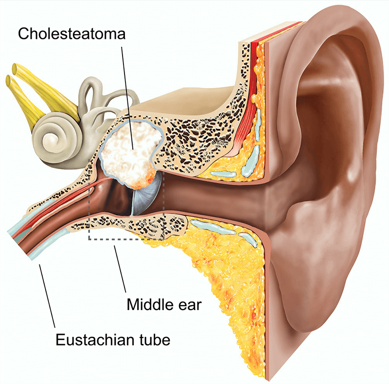

What Causes Cholesteatoma?
Cholesteatoma occurs when recurrent ear infection and eustachian tube function is inadequate. The Eustachian tube provides ventilation of the middle ear. The Eustachian tube provides air passage through the nasal cuff to compensate for middle ear pressure. In cases such as allergies, colds and sinusitis, the Eustachian tube cannot function and vacuum (negative pressure) occurs. Due to inflammation, the tapered eardrum is withdrawn by this vacuum. In this pouch, skin growth called cholesteatoma starts.
What Are The Symptoms Of Cholesteatoma?
The symptoms of cholesteatoma are initially mild. However, with the growth of the cyst, the symptoms begin to get worse and more severe. One of the symptoms of cholesteatoma is smelling discharge. At the same time, with the increase in intra-ear pressure increases discomfort, but can lead to deafness. As the cyst grows, the face begins to be affected. If the face starts to be affected, problems such as weakening of facial muscles or facial paralysis may be unavoidable. The growing cyst causes dizziness.
- Ear infection
- Bad smell in the ear
- Temporary or permanent hearing loss
- Tinnitus
- vertigo
- Slimming facial muscles
- Inner ear cavity
- Cyst spread to the brain
- Meningitis
- Brain abscess
- Continuous ear drainage
Cholesteatoma Treatment
Thorough cleaning of the ear should be ensured before cholesteatoma treatment is initiated. Antibiotic and drip treatment should be applied with the cleaning of the ear. Thus, while reducing inflammation is prevented at the same time spread.
At this time, the size and degree of cholesteatoma are determined. Surgical intervention may be required in large and risky cholesteatoma cases. Surgical interventions are performed under general anaesthesia, while the removal of cholesteatoma and a healthy ear structure is the aim of the surgery. The other purpose of the surgery is to improve the quality of hearing by improving.
However, in very advanced cases of cholesteatoma, the hearing may not be completely corrected. Cholesteatoma surgery can be performed on the day the patient visits the doctor and the patient can be discharged on the same day. However, depending on the degree of cholesteatoma, the patient may need to be supervised and treated with serum and antibiotics.
Vertigo (Dizziness)
Vertigo; Latin comes from the verb “to return” and is dizziness caused by a problem in the balance system. Vertigo is defined by patients turning around themselves or oneself. If there is only one lightheadedness without rotation, it would be more accurate to define it as dizziness rather than Vertigo.
Vertigo can be severe enough to turn patients’ daily activities into nightmares, cause the patient to go to bed and cause pain that she cannot even open eyes, but it can also cause a feeling of slippage and blackout from time to time.


Some methods used in the diagnosis of vertigo are:
- Pure Sound Audiometry
- Skull x-ray
- Tomography
- VNG-guided bilateral caloric test
- VEMP
- Posturography
In the treatment of Vertigo: in addition to medication, Semont, Epley and Log Roll (barbecue) manoeuvre, surgical treatments are also applied.
What are The Diseases that Cause Vertigo?
Vertigo is classified as Peripheral and Central Vertigo according to its origin.
-
Peripheral Vertigo
Peripheral Vertigo is caused by a disorder in the inner ear or vestibular organs.
The most important causes of Vertigo from the inner ear are:
- Benign Paroxysmal Positional Vertigo (BPPV)
- Meniere’s Disease
- Vestibular Neuritis
- Perilymph Fistula in Ear
The causes of the vestibular nerve are:
- Meningitis
- Syphilis
- Viral (Mumps)
- A tumour (Acoustic Neurinoma, Meningioma)
Conditions such as cold and trauma that affect the inner ear and some types of antibiotics may also cause this type of Vertigo.
-
Central Vertigo
Central Vertigo is caused by a problem in the balance centres in the brain. Status; speech disorders, double vision, swallowing disorder. The severe imbalance is common.

How is Vertigo Diagnosed?
History of the disease is very important in the diagnosis of vertigo. vertigo; your doctor will diagnose you according to the nature of the dizziness you are experiencing.
How Vertigo is Treated
After the patient is diagnosed with Vertigo, specific treatment for the cause should be planned. In the treatment of vertigo, ENT, neurology, internal medicine and physical therapy evaluations may be necessary. The proposed treatment will be associated with the diagnosis. It is recommended that a control examination be performed at least once a year and regular use of drugs.
The treatment covers a wide range of things, from simple actions your doctor will have during the examination, to surgical treatment.
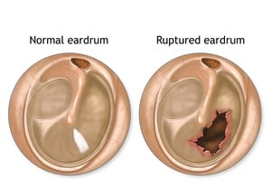
Perforation of the Eardrum
The eardrum is a thin membrane that separates the middle ear from the outer ear canal and is a very important component of the human sensory organ. Perforation of the eardrum occurs as a result of rupture or perforation of this membrane.
When the eardrum is punctured, hearing is reduced and sometimes there is a discharge from the ear. A tearing and ringing sound can be felt in the ears, but usually, no pain occurs. Perforation of the eardrum may occur suddenly after a sensation similar to thunder. In some cases, you may experience sharp pain in your ears and the pain may disappear suddenly.

Causes of Tympanic Membrane Perforation
Various activities in daily life can cause pressure changes in the ear and perforation of the membrane. This condition, known as barotrauma, results from the fact that the pressure inside the ear is different from the outside.
Conditions that may cause perforation of the eardrum;
- Scuba diving
- Airplane trip
- Exposure to shock waves
- Driving at high altitudes
- Injury and trauma
- Sudden pressure changes
- Insertion of foreign bodies into the ear
- Inflammation of the middle ear
- Hot or acidic liquids entering the ear canals
- Car accidents
- Sports injuries
- Falling on the ear
- A hard blow to the ear
- Explosion near the ear
Treatment of Eardrum Perforation
In general, perforations in the eardrum are quickly repaired by the body. This repair is the same speed for both adults and children. Surgical intervention may be required in the eardrum holes that do not close itself within six weeks.
The neglected hole in the eardrum can cause inflammation in the middle ear and related chronic disorders, hearing loss.
Different surgical methods can be used for the treatment of non-occluded eardrums, although drug treatment and paper closure have been performed with a patch method (3 44 patches may be required to completely close the hole). The common point of these methods is; is the closure of the membrane with a tissue to improve puncture. This process is called “myringoplasty.”
Eardrum Holes And / Or Persons In The Stage Of Treatment
- Water should not have seeped into the ear (when bathing or washing your head, a piece of cotton wool covered with petroleum jelly may be put in the ear, while swimming, a tight bathing cap should be worn over the cotton. Or earplugs should be used).
- Strong blowing should be avoided and runny nose should be withdrawn. If it is necessary to blow the nose, it should be done without closing the other nostril.
- If there is an ear discharge, it should be cleaned without inserting anything.
- The drug should be used if there is an ear discharge or if it has started.
- The ear should be protected from trauma.
Fluid Accumulation in the Middle Ear
What is Fluid Accumulation in the Middle Ear?
Normally a small amount of fluid is produced in the middle ear. This fluid usually flows from the ear through the eustachian tube that connects the middle ear to the back of the nose.
Accumulation of fluid in the middle ear occurs when fluid accumulates in the area behind the eardrum. This fluid can cause problems in children. This usually does not cause pain. It most often occurs after otitis media, but it can also occur without previous infection. It tends to heal itself, but if your child has certain symptoms, then you will need to see a doctor.

Accumulation of Fluid in the Middle Ear and Risk Factors
The reasons are:
- Eustachian tube blockage
- Cold
- Ear infection (otitis media)
- Cleft lip
Approximately 10% of children remain fluid in the ear 3 months after infection.
Risk factors include:
- Frequent ear infections
- Frequent colds
- Cleft palate
Symptoms of Fluid Accumulation in the Middle Ear
If your child has certain symptoms, he or she should be examined by a doctor. Symptoms include:
- Hearing problems
- Reaction or carelessness
- Backwardness
- Pulling or pulling the ear
- Muffled noises
Fluid Accumulation Treatment in Middle Ear
This condition usually resolves by itself within 4-6 weeks. Antibiotics are not needed unless your child also has an upper respiratory tract infection.
If the condition lasts longer than 2 or 3 months, it may be necessary to insert a tube into your child’s ears. This procedure is performed under general anaesthesia. A surgeon places a small drainage tube into the eardrum to aid in the discharge of fluid.
Most of the time, the fluid in the ears goes by itself. As your child grows, the eustachian tube grows; this means that the liquid will have more space to flow from the ear.
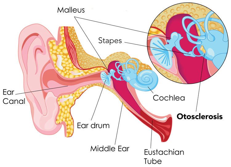
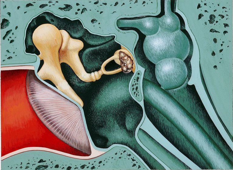
Otosclerosis (Calcification of The İnner Ear)
Otosclerosis, known as inner ear calcification, is a common cause of hearing loss. In otosclerosis, solidification occurs as a result of structural calcification in the wall where the stirrup ossicle is adjacent to the inner ear and movement restriction occurs in the stirrup bone. Consequently, sound waves cannot be adequately transmitted to the inner ear fluids and hearing loss, called the conduction type, occurs. In this case, the inner ear is intact. Only voices cannot be transmitted. However, in the latter stages of the disease, this calcification may also affect the inner ear wall and neural type hearing loss may also occur. Otosclerosis may be a genetic disease.

What Are The Symptoms of Otosclerosis?
The main symptom of otosclerosis is slow progressive hearing loss that can affect one ear or both ears. The extent of hearing loss may vary with the severity of calcification. The disease is usually seen in the range of 15 to 45 years. It is more common in women than in men. During pregnancy, the disease may enter a period of rapid progression. In addition to hearing loss, tinnitus, dizziness and balance problems can also be seen in patients.
How is Otosclerosis Diagnosed?
Physical examination is normal in the diagnosis of otosclerosis; that is, the eardrum is seen as normal. The hearing test is required for the diagnosis of the disease. With this test, the degree of hearing loss and its origin can be predicted. Imaging methods (tomography or magnetic resonance) do not indicate otosclerosis but may be preferred to differentiate other ear diseases. Otosclerosis does not affect the general health of patients and is often a curable disease.
What are The Treatment Options in Otosclerosis?
In patients with a pre-diagnosis of otosclerosis, a treatment plan is made according to the severity of the disease and the preferences of the patient. The patient can be followed up with hearing tests in cases where he is new and does not affect the patient clinically. For patients whose hearing loss affects their social life, surgery or rehabilitation with hearing aids may be preferred. There are also some drug therapies, such as sodium fluoride, which slow down the progression of the disease, but these drugs are not the most preferred treatment modality.

What is Otosclerosis Surgery?
The method called ‘‘Exploratory tympanostomy’’ is preferred for surgical treatment of the disease. During surgery, the middle ear and ossicles are observed behind the eardrum. During the operation of the stirrup, bone movement is controlled with the help of a tool. If the movement is found to be restricted, otosclerosis is definitively diagnosed. During the surgery, the stapedotomy/stapedectomy technique is used to remove the stirrup bone, which is restricted in movement, and a prosthesis (Teflon or metal) is placed between the anterior bone and the inner ear to transmit sound waves.
Complications of the surgery are very rare. There may be dizziness for a few days after surgery, but this is a temporary condition. There may be continued or even worse hearing loss.
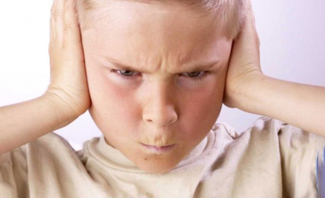
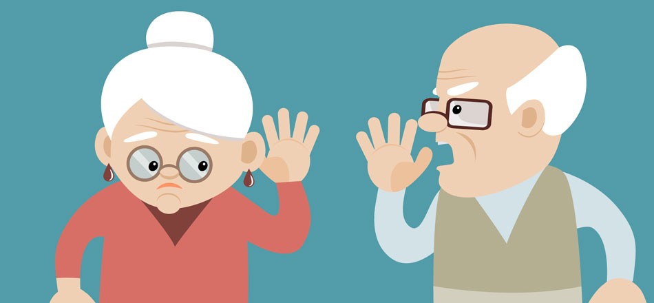
Hearing Problems
Our hearing is one of the most important tools in our perception of the outside world, sometimes due to congenital and sometimes discomfort caused problems. The existence of a hearing problem, especially during childhood, where development is the fastest, adversely affects the child’s social life and learning ability.

What Causes Hearing Loss?
The severity of hearing loss varies from mild to permanent. Some hearing problems can be diagnosed by examination, while others may require multiple tests.
Hearing losses are generally divided into 3.
- Conductive hearing loss due to external and middle ear diseases
- Sensorineural hearing loss due to the inner ear, hearing nerve and brain-related diseases
- Mixed (mixed) hearing loss due to damage in more than one region of the auditory tract
The causes of hearing loss according to parts of the ear are:
Outer Ear: Earwax, foreign body, absence of auricle or congenital deformity in the outer ear, congenital obstruction of the external ear canal or ear-shaped disorder, external ear inflammation or tumour
Middle Ear: inflammation in the middle ear, low pressure, calcification of the middle ear, tumours in the middle ear, torn or punctured eardrum
Inner Ear: The hearing nerves in the cochlea may be damaged due to ageing and exposure to high noise. Inheritance can make one more susceptible to these changes. This type of hearing loss is also known as permanent hearing loss. Also, inflammation of the inner ear, Meniere, tumours in the auditory nerve can cause hearing loss.
What are the Risk Factors Causing Hearing Loss?
Age: Prolonged exposure to sounds can damage the hearing nerves. This increases the risk of hearing loss.
Genetic Heritage: Your genetic predisposition increases your likelihood of hearing loss.
Profession: Persons working in noisy environments (eg factory, construction site) may experience hearing problems due to long exposure to loud sound.
Some Medications: Antibiotics and some medications used in chemotherapy may cause damage to the inner ear. Overdose may cause temporary hearing problems such as aspirin, painkillers, diuretics, tinnitus or hearing loss.
Some Diseases: Diseases such as meningitis with high fever can damage the cochlea and cause hearing loss.
Because the symptoms of hearing loss are not always clear, people tend to ignore this problem. Therefore, many people do not go to the doctor before hearing loss progresses. However, the sooner the problem is detected and treated, the sooner the hearing loss can be prevented and the right treatment can be applied.
Signs and symptoms of hearing loss include:
- Difficulty in the perception of speech and other sounds,
- Inability to perceive background sounds and words, especially in noisy and crowded places,
- Asking people to speak slowly, clearly and loudly,
- The need to turn on the television or radio,
- Starting to avoid speaking and some social situations
How is Hearing Loss Diagnosed?
To determine the cause of hearing loss, the ENT specialist will first examine the ear and look for any problems with the outer ear or membrane. From the appearance of the eardrum, it will be understood how the middle ear looks like. If this examination does not reveal a problem, hearing loss is thought to be caused by the inner ear and many tests, especially audiometry, are performed.
In audiometry, that is, the type, degree, and frequency of hearing loss are determined. Tympanometry may be required to measure middle ear pressure, stapes reflex to measure calcification of the ear ossicles, and computed tomography (CT) or (MR) testing may be necessary to determine the cause of hearing loss, especially in the inner ear.

How Are Hearing Problems Treated?
The treatment of hearing problems varies according to the cause and severity of the problem. Treatment options include:
Cleaning Earwax: Ear blockage caused by earwax accumulation is a treatable condition. Your doctor may remove earwax for treatment by softening.
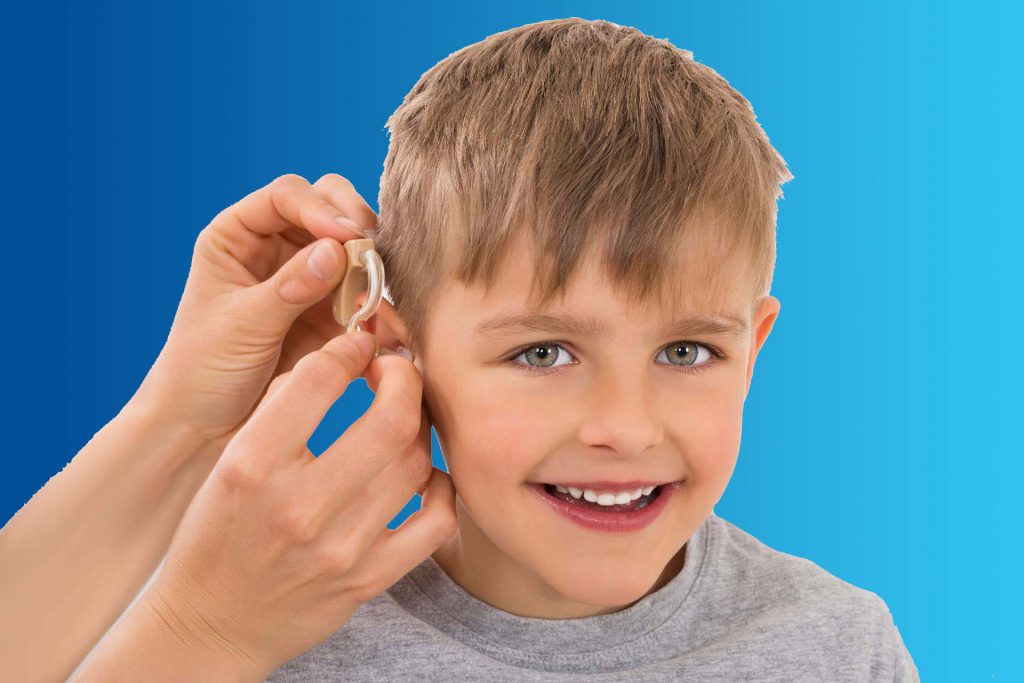
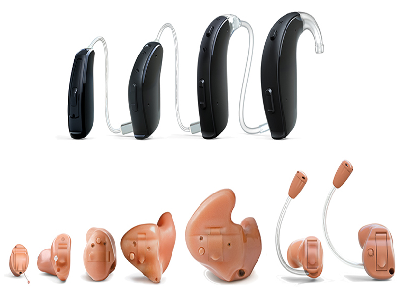
Hearing Aids: If hearing loss is caused by damage to the inner ear, hearing aids can make hearing sounds stronger and easier. The structure of the hearing aids varies according to the needs of the person.
Cochlear Implant: Cochlear Implant is a medical device used to eliminate problems in the inner ear. Hearing aids increase the volume of sound; The cochlear implant performs the tasks of the cochlea, which has problems in sending sound signals to the brain. It stimulates the hearing nerve and is perceived as sound by the brain.
What are The Psychological Effects of Hearing Loss?
Hearing loss has a significant impact on people’s quality of life. For example, the common problems of elderly adults with hearing loss are:
- Depression
- Anxiety
- The thought of others being angry at him for not hearing.
These thoughts negatively affect both social and work life.