Horner Syndrome
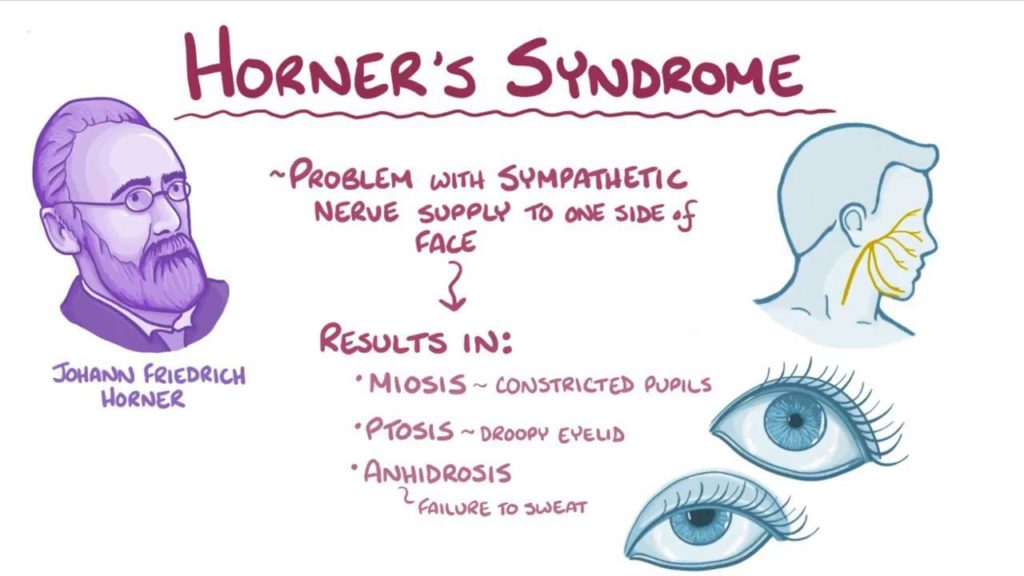
Horner syndrome is a neurodegenerative eye disease. In this syndrome, which develops clinically in sympathetic nervous system damage, lesions occur in the upper eyelid, pupil, facial sweat glands and fibers that go to the vessels.
Especially in children, it causes results such as different iris of the two eyes, called heterochromia. In general, the eye color in which sympathetic innervation develops is lighter..
What Causes Horner Syndrome?
- Apex tumor: Damage to nerve fibers, usually with the progression of lung cancer to the stethate ganglion,
- Anomalies in nerves with intracranial lesions,
- Spinal cord injuries,
- Problems occurring in the neck sympathetic nerve network,
- Serious throat infections,
- Congenital causes (head and neck trauma at birth)
- Nervous system cancers
What are the Symptoms?
- Pupillary constriction in the eye where the discomfort develops,
- Low upper eyelid called ptosis,
- Reverse ptosis, that is, elevation of the lower eyelid,
- Iris discoloration in the eye affected by Horner’s syndrome,
- The end of the ability to sweat on the face, usually a part of the face does not sweat.
- Inward pulling of the eyeball
Which Tests Are Used to Detect Horner Syndrome?
Tests to support clinical findings should be at a level to prove the presence of Horner’s syndrome. First of all, pharmacological tests with eye drops used by the ophthalmologist will be sufficient.
Especially when this disease is diagnosed in children, some tests may be required on the abdomen, chest, neck and neurological aspects of the specialist patient.

